
Cervical Arthroplasty
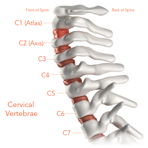
The cervical spine is the upper part of the back between the base of the head or skull and the thoracic spine, which begins around the of the shoulders. The cervical spine is also known as the neck. It is made up of seven bones or vertebrae (C1 to C7) that are stacked on top of each other. The cervical spine supports the head and provides for movement of the neck. The neck moves from side-to- side (rotation), forward (flexion), and backward (extension).
In between each bone (vertebra) are spinal discs. These discs act like shock absorbers to cushion and protect the bones and allow for motion. Each spinal disc is made up of two parts. The inner part is a jelly-like substance and is called the nucleus pulposus. The outer part is a stronger rubber band-like structure called the annulus fibrosus. The annulus protects and holds the nucleus in place. The nucleus is the cushion of the spine. Additionally, there are muscles and ligaments running alongside the bony spine to help with support and movement.
A canal runs down the middle of the bony spine; the spinal cord sits within this bony canal and extends from the brain to your tailbone. The bones protect the spinal cord. There are small openings, or foramina, on the sides of the bony canal that let the spinal nerves leave the spinal cord and go to different parts of the body. The spinal cord carries messages between the brain and the rest of the body to provide movement and sensation (feeling) to the entire body. In the neck, nerves from C1 to C7 branch out and go to the shoulders, arms, and hands to provide movement and feeling.

A canal runs down the middle of the bony spine; the spinal cord sits within this bony canal and extends from the brain to your tailbone. The bones protect the spinal cord. There are small openings, or foramen, on the sides of the bony canal that let the spinal nerves leave the spinal cord and go to different parts of the body. The spinal cord carries messages between the brain and the rest of the body to provide movement and sensation (feeling) to the entire body. In the neck, nerves from C1 to C7 branch out and go to the shoulders, arms, and hands to provide movement and feeling.
Cervical Disc Degeneration
As a person ages, it is common for the spinal discs in the neck (cervical spine) to slowly undergo wear and tear. These discs, which are the cushions between the bones (vertebrae), tend to dry out and lose height over time—a process known as cervical disc degeneration or degenerative disc disease. As the discs become thinner and less flexible, the neck bones move closer together. This change may increase stress on the surrounding joints, ligaments, muscles, and spinal nerves.
As cervical spinal discs degenerate, the body may respond by forming bone spurs (osteophytes) along the edges of the vertebrae to help stabilize the spine. While these bony growths may limit excessive motion, they can also reduce the space within the spinal canal or the openings around nerve roots. This narrowing may cause pressure or compression of the spinal nerves or the spinal cord. In turn, this pressure may cause symptoms such as neck pain, neck stiffness, lack of neck motion, or tingling, numbness, or weakness in the shoulders, arms, or hands.
Continued disc degeneration may cause a cervical disc to push into (herniate) the nerve structures, putting
pressure on the spinal nerve or the spinal cord. Cervical disc herniation may be felt as tingling, numbness,
or as a shooting pain in your arm or hand, or perhaps as muscle weakness; doctors call this radiculopathy.

Taken together, these changes to the cervical spinal discs (disc wear, herniation, and bone spurs) are called cervical degenerative disc disease.
Symptoms of Cervical Disc Degeneration
Another condition called cervical myelopathy may also occur due to disc degeneration. This condition
results when the spinal cord becomes compressed. This compression or pressure on the spinal cord may be
caused by various spinal changes, such as a collapsed disc or a bone spur protruding into the spinal canal.
Cervical myelopathy may interfere with the spinal cord’s ability to transmit signals between the brain and the rest of the body. Symptoms of cervical myelopathy may include difficulty with the ability to button a shirt, unsteadiness when walking, and muscle stiffness or weakness in the arms or hands.

Diagnosing Cervical Disc Degeneration
Diagnosing cervical disc disease involves a complete evaluation by a spine surgeon. The surgeon will evaluate your symptoms, take a detailed medical history, do a physical exam, and do imaging studies, like X-rays or Magnetic Resonance Imaging (MRI).
During your physical exam, your surgeon will examine your posture, your neck movement, your muscle strength, your reflexes, and the areas where pain, tingling, or weakness are felt.
Based on this exam, if cervical disc degeneration is suspected, your surgeon may order diagnostic imaging to confirm the diagnosis. X-rays are used to look at the alignment of the spine and measure cervical spine disc height. An MRI provides detailed pictures of the spinal discs, nerve roots, and spinal cord. The MRI allows the spine surgeon to see disc or bone changes, like herniated discs, bone spurs, or the narrowing of the spinal canal.
Cervical Disc Degeneration Treatment Options
Treatment options for cervical degenerative disc disease range from non-surgical therapies (e.g., bed rest, physical therapy, and anti-inflammatory medications) to surgical intervention for nerve or spinal cord compression.
In most cases, conservative, non-surgical treatments are the first line of patient care. The goal of these treatments is to reduce symptoms, improve physical function, and help patients return to their daily activities.
If symptoms do not improve with non-surgical care or if there is significant nerve or spinal cord compression, surgical intervention may be recommended. The primary goal of surgery is to relieve pressure on the nerve or spinal cord. This procedure is known as decompression. Decompression involves removing the damaged disc and any bone spurs that are contributing to the compression.
After decompression, the cervical spine surgeon may perform one of two surgeries in the neck: anterior cervical discectomy and fusion (ACDF) or artificial disc replacement (ADR).
In ACDF, the damaged disc is removed from the front of the neck, and a special spacer called an interbody device is inserted into the disc space to replace the disc that was removed. A metal plate and screws are often affixed to the spine to stabilize (support) the spine while the neck bones (vertebrae) fuse together. An ACDF limits motion in that part of the neck and places additional pressure on the remaining spinal bones.
For patients who meet specific criteria, artificial disc replacement may be an option. After the diseased disc is removed, a device such as the Synergy Disc can be placed (implanted) in the disc space. The artificial disc replacement surgery is designed to restore normal disc height while preserving motion at that spinal segment, while allowing adjacent spinal segments to retain natural motion.
Cervical Spine Artificial Disc Replacement Spine Surgery
Artificial disc replacement surgery is a type of spine surgery that helps people with neck or back pain (myopathic symptoms) or pain/numbness/ tingling in the arm (radicular symptoms), caused by a diseased or damaged spinal disc. During artificial disc replacement surgery, a surgeon removes the diseased or damaged disc and replaces it with an artificial disc. An artificial disc is designed to restore height, movement, and function like a healthy, natural disc. It allows the spine to bend and move in contrast to traditional spinal fusion surgery (ACDF), where the bones are locked, or fused, in place.

Cervical (neck) artificial disc replacement surgery is performed in several steps. First, the cervical spine surgeon makes a small incision or cut across the front of the neck near the diseased or damaged disc. After gently moving aside muscles and soft tissue, the surgeon removes the damaged spinal disc and osteophytes. The space where the disc was then prepared for the new cervical artificial disc. Next, the surgeon carefully places the artificial disc into that disc space. To make sure it’s in the right place, the surgeon takes a few Xrays (fluorographs). Once disc placement is optimal, the incision in the neck is closed with stitches.
